|
Nathan Grant, Eugueni Matveev, Amanda S. Kahn, Sally P. Leys
doi.org/10.1016/j.marenvres.2018.02.020 Sponges filter water to capture and eat bacteria by using an intricate filtration system. But what happens when something unwanted, like sediment, gets into their filter? Glass sponge skeletons are too rigid to “sneeze out” the particles (that's what demosponges do); instead, they just stop pumping water to avoid taking in any more irritants. Although this response by glass sponges was already shown in the lab, in this paper we showed for the first time that it also happens in the sponge’s natural environment. We conducted our research off the coast of British Columbia, where vast expanses of reefs spread as far as our submersible could see. By placing flow probes in the top opening of the sponge (called the 'osculum'), we were able to measure how much water they were pumping. Then, we scooped up some sediment from the seafloor, and dispersed it around to increase the sediment levels around the sponge. Based on our analysis of the flow, we found that sponges do indeed show more “arrests” in feeding when sediment levels are increased. This kind of research is important because human activities on the seafloor (such as bottom trawl fishing) kick up massive clouds of sediment that can travel for miles. If our activities affect the health of the reefs, we could be putting this unique ecosystem in danger. |
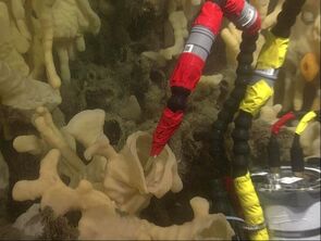
A flow probe inside a the osculum of Aphrocallistes vastus.
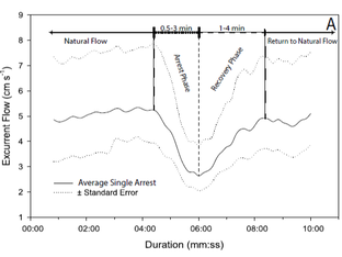
A diagram of the arrest of a sponge feeding current.
|
|
Leys, S.P and Mackie, G. O.
doi:10.1038/387029b0 Even without a nervous system, sponges can respond to their surroundings. When touched or under high sediment levels, glass sponges briefly stop pumping water; this behavior is called a “filtration arrest”. Seemingly simple, a coordinated whole-body arrest means that sponges have some nerve-less signaling system between different parts of the body. Based on the speed of response it was suspected that the signal could be electrical; but, sponge tissues are fragile, so it's difficult to place electrodes and measure electrical pulses without damaging the sponge. To avoid this, we mushed up tissue from the sponge Rhabdocalyptus dawsoni, got the tissues to re-form into a sizeable lump, and grafted this lump onto a sponge. We could then place electrodes onto the sponge without causing damage. By stimulating the sponge while measuring the flow and electrical current, we saw that every filtration arrest was preceded by a wave-like electrical impulse. The wave pattern was similar to one we see in nerve cells: an action potential. But how can an action potential travel without nerves? Three quarters of R. dawsoni tissue is composed of a “syncytium”: a mass of fused cell bodies without cell membranes. It is this lack of cell membranes that we think allows for an action potential to freely travel at the speed of up to 0.3 cm per second throughout the sponge. Could an action potential in a nerve-less multicellular animal be a hint towards an important step in the evolution of nerve cells? |
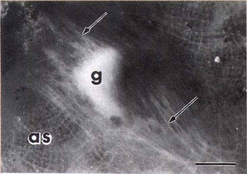
Grafted clump of aggregated R. dawsoni tissue (g) on the atrial surface (as) of the sponge. Arrows show sponge cytoplasm attached to graft.
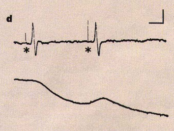
Sponges were stimulated (asterisks), after which an action potential was recorded (top line) and the flow velocity decreased (bottom line).
|
|
Danielle A Ludeman, Nathan Farrar, Ana Riesgo, Jordi Paps & Sally P Leys
https://doi.org/10.1186/1471-2148-14-3 To get rid of unwanted particles in the body, some sponges elicit their famous “sneeze” behaviour. But, not having nerve cells, how can they sense these irritants and respond? One contender for a sensory mechanism are non-motile "primary" cilia. These small hair-like "antennae" play an important sensory role in organisms from unicells to humans. We wanted to find out if sponge cells in the osculum have primary cilia, and if so, whether they play a similar sensory role as in other animals. To confirm that primary cilia were present in sponges we used a combination of electron microscopy and antibody labeling. And indeed, we saw two cilia in the shape of a ‘V’ in the center of each sponge cell (top images). Cross sections of cilia resembled those of other animals, and high-frequency time-lapse microscopy showed the cilia did not move; we concluded that sponges have non-motile cilia comparable to other animals. The next step was to confirm that the cilia did, in fact, have a sensory role in sponges. We treated live sponges with three chemicals that stop the function of cilia in other animals. Sponges that were treated with chemicals had significantly reduced “sneezing” responses, confirming that primary cilia likely play a sensory role in sponges just like in other animals. This research furthered our understanding into how, even without nerves, sponges can sense the world around them. |
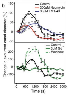
Three chemicals (Neomycin, FM1-43, and Gd3+) that inhibit primary cilia in other animals also decreased the sneeze response.
|
|
The transition from individual cells to multicellular animals is a crucial and fascinating point in evolution. Yet, we know very little about the animals with the highest chance at the title of “first animal”: sponges and ctenophores. Humans, as Bilaterian* animals, tend to view the animal world through a Bilaterian lens. This means we are biased, and tend to look for traits and genes resembling ours in other animals. But sponges and ctenophores are so vastly different from us that this approach just doesn’t make sense. Because there are so few traits and genes that resemble ours in these animals, it leaves a large portion of the sponge and ctenophore biology un-researched, and thus ‘hidden’. In this review, we argue that the assumption that sponges and ctenophores are "simple" animals is a reflection of our lack of understanding, not actual simplicity. Sponges, despite lacking nerves and typical germline layers, have sensory systems and tissues that are so different from ours they would not be recognized by conventional methods. Similarly, while ctenophores bear a superficial resemblance to jellyfish (cnidarians), closer inspection of these similarities shows they have very different underpinnings. We argue that for us to untangle the complicated web of early animal evolution, we must not group organisms into “simple” or “complex” based on our biased Bilaterian perception of complexity. Instead, we must research these animals not in comparison, but in their own right. *meaning, we have a mirror like symmetry down the center of our bodies. |
Because we have a bilaterian bias, we are missing a large portion of non-bilaterian specific biology.
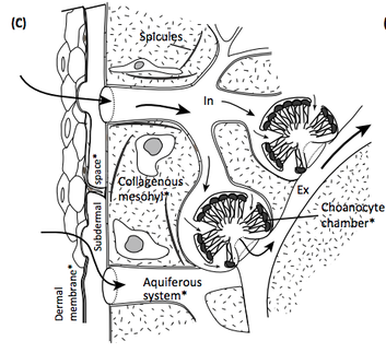
Seemingly simple, sponges have a complicated morphology we are just starting to understand.
|
|
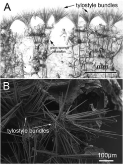
Lauren K. Law, Henry M. Reiswig, Bruce S. Ott, Neil McDaniel, Amanda S. Kahn, Keenan C. Guillas, Curtis Dinn, Sally P. Leys
The reef-building glass sponges off the coast of North America grow a rigidly latticed skeleton of silica “glass”. The skeleton allows the sponges to tower above the seafloor. The folds and crevices within them are used as a home by countless species. In this way, the reefs increase the biodiversity around them, and in 2017 the Hecate Strait Queen Charlotte Sound Glass Sponge Reefs in northern British Columbia were designated as a Marine Protected Area. Growing tall is an advantage to the sponges too; higher tidal currents bring them more food. But not all sponges can grow an upright skeleton. Sponges in the genus Desmacella, for example, grow as a ‘crust’ on top of the glass sponges to reach the higher flows. Like a SciFi zombie-sponge, they take over they skeleton of glass sponges to use as their own. Very little is known about Desmacella species, but estimating their prevalence is important to accurately assess biodiversity in these ecologically important areas. Using the Remotely Operated Vehicle “ROPOS” we took photos along the seafloor, and collected Desmacella specimens. We even used samples collected by Citizen Scientists (thank you to Bruce and Neil), and old museum samples. Desmacella spp., we found, were found in higher densities when either dead and live glass sponges were present. In some locations they constituted nearly 20% of the sponge proportion. By analyzing the skeletal “spicule” components of our Desmacella specimens we found a new species which we named Desmacella hyalina, after the greek word huálinos, meaning “crystal glass”. We suggest that the diversity of sponges can be hidden in plain sight. To accurately assess how the species number and composition is changing over time, we need to put in a lot of work into better understanding the species that are present. |
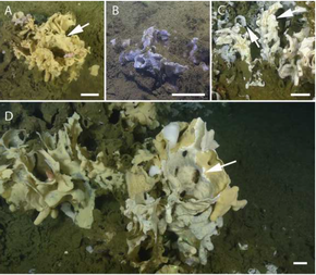
The different desmacella sponge colour morphs (arrows) blend in with glass sponges.
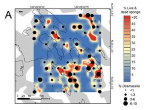
The desmocella concentration is higher in areas where there are more live/dead sponges.
Some of the "glass spicules" in the new Desmacella hyalina species.
|
